 Baby ultrasound is a common, safe and fun way to see your unborn baby’s development. It is especially helpful for women who may be concerned about a medical condition that could affect their baby. It can also be used to estimate due date and calculate the size of the fetus. It is also a valuable tool for doctors to look at the fetal organs and the baby’s heart, lungs and kidneys. Having a baby ultrasound reveals the development of the fetus, but most scans show that the baby is developing normally. It can also help doctors diagnose certain medical conditions that require additional treatment or monitoring. During the scan, the healthcare provider spreads a clear water-based gel on your belly and pelvic area. This helps the probe to transmit sound waves properly. Then the health care provider moves a small wand (transducer) over the area to capture images on an ultrasound machine. The technician may take some measurements of the area and make notes on a computer screen. You may hear a buzzing noise or feel pressure as the transducer is moved over your body, but this is nothing to worry about. When the doctor has finished the scan, you will be given some printed pictures to take home. These pictures will usually show white or gray areas showing bone and tissue, while dark areas show liquid, such as amniotic fluid. If you have a problem with your bladder, you may be asked to drink a lot of water before the scan. This helps the sonographer use a 'porthole' to get a clear view of your uterus and other structures. You will then lie on an examination table or bed and the healthcare professional will apply a gel to your abdomen and pelvic area. This gel helps the sound waves travel through your body properly and produces clear images of your baby on the ultrasound monitor. This is a simple, painless procedure and takes less than 30 minutes. The healthcare professional may ask you to hold your breath or move slightly as they move the transducer over your body. You can also find some hospitals that offer a special 3-D or 4d ultrasound at pregnancy checkups, which can give you a better view of your baby's face and movement before birth. It can help doctors to identify abnormalities such as cleft lip and palate, spina bifida, and other congenital conditions. The 3-D and 4-D ultrasounds are generally safe because they use non-ionizing radiation, unlike X-rays, which can cause problems for you or your baby. But it is important to note that these types of exams aren't recommended by the American College of Obstetricians and Gynecologists, the Food and Drug Administration or the American Institute of Ultrasound in Medicine. Some stores or fetal portrait studios offer "keepsake" 3-D or 4-D ultrasound pictures or videos for parents-to-be that aren't done by trained professionals. Experts warn against these non-medical ultrasounds, as they can be inaccurate or harmful to you and your baby. To familiarize yourself more with this topic, it is best that you check out this post: https://en.wikipedia.org/wiki/Obstetric_ultrasonography.
0 Comments
5/4/2023 0 Comments What is a Baby Ultrasound?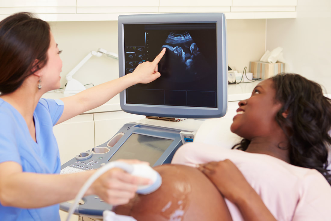 Ultrasound is an imaging procedure that uses sound waves to produce pictures of your baby. It is used to check your pregnancy, screen for problems, and give you an idea of how big your baby is. It is an important medical examination and usually shows that everything is normal. The health of your baby is monitored and if there are any problems identified they will be treated as soon as possible. A baby ultrasound is a safe and quick test that will show your baby's position, size and growth. It also checks the development of your baby's heart, lungs and brain. During your scan you will be asked to drink plenty of fluids and not urinate for a while so that your bladder is full. You will be put on a bed or exam table and a technician (sonographer) will spread a gel over your abdomen, which makes sure the sound waves travel properly. They will then place a small wand over your belly and move it to capture images of your baby on the ultrasound screen. An ultrasound can also help your provider determine your due date and the number of weeks you are pregnant. You will then be given a printout of the image. This will be very useful in deciding when to deliver your baby. There are many types of ultrasound that your doctor can do to see your baby. Some of these include - Transvaginal Scans – This type is done when you are in the first or second trimester, between weeks 6 and 7. A narrow ultrasound wand, called a transducer, will be gently inserted into your vagina and moved around inside your uterus to capture an image of your baby’s organs. It may feel uncomfortable, but it should be painless and safe for your baby. Doppler – This type of ultrasound is also used to look at your baby’s heart rate, and it can identify abnormalities that are caused by certain blood vessels, such as the umbilical cord. This will give your doctor more information to help make a diagnosis if you have any concerns. 3D and 4D - These types of ultrasounds are a new development in ultrasound imaging that can be helpful to doctors in identifying specific fetal abnormalities and congenital diseases. These can be done in ultrasound clinic london and are recommended for certain conditions that require the doctor to have more detailed and lifelike images of the fetus. Abnormalities that are found by these ultrasounds can cause women to have a lot of worries about their child, and they might need to make difficult decisions about whether to abort or continue their pregnancy. It's important to talk to your doctor about these issues before you have the scan so that you can be fully informed and understand what to expect. During the scan, you will be asked to change into a gown and lie down on a table. The room will be dark so you should be able to see the images clearly on the ultrasound screen. Check out this post for more details related to this article: https://en.wikipedia.org/wiki/Ultrasound. 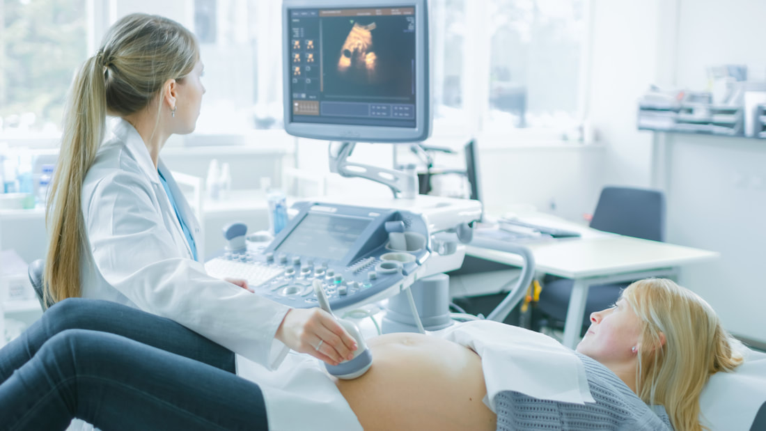 A baby ultrasound, also called a sonogram or fetal ultrasound, is an imaging technique that uses sound waves to see your unborn child inside your womb. It is performed by a healthcare provider or medical technologist and can be used to detect and diagnose certain health problems and developmental abnormalities. It is a painless procedure and typically takes 30 minutes or less to complete. During your pregnancy ultrasound 3d procedure, the technician applies a gel to your abdomen (tummy) and moves a probe over the area. The sound waves reflect off the fetus and back to the transducer, which sends signals to a monitor that creates black-and-white images. You may be asked to lie on one side during the scan or to turn on both sides if images are needed from different positions. The basic anatomy scan - This type of ultrasound is done at about 12 weeks of pregnancy and uses sound waves to examine the baby's head, body and bones. It can help your doctor determine how far along you are and estimate your due date. It can also help identify any issues, such as multiple births or a placenta that is not growing normally. You will be asked to lay on your side and a thin gel will be applied to the surface of your tummy. This is to allow better contact between the scanner and your skin. Then the ultrasound probe is moved over your tummy and pictures are taken, which are sent instantly to a computer screen. The images are very clear and show a detailed view of your baby's body. At six weeks, an embryonic heart can be seen on the ultrasound screen. The technician can also view the fetus' brain and its internal organs, as well as the mother's uterus and the placenta. In some cases, a gynecologist can assess your baby's sex at this ultrasound by looking at the position of their genitals. However, this is not always possible. If you don't want to know, let the gynecologist know beforehand. If you are having a transvaginal ultrasound, the ultrasound hamilton practitioner will insert a small wand-like probe into your vaginal cavity. The wand transmits high-frequency sound waves that bounce off of the fetus and return to the device. The images are then converted to black-and-white pictures that the doctor can view on a screen. Your baby's size - This ultrasound is often done at the beginning of your third trimester to check that your baby is still on track for growth. The scan can also measure your baby's limb and finger lengths. If your baby is too short, this could indicate that they are not getting enough nourishment during the first few months of their life. During the ultrasound, your doctor can measure the width and length of your baby's rib cage and the bi-parietal diameter of the bones in your pelvic region. This information can be useful in predicting your baby's future weight and height. You can also choose to have a nuchal translucency scan, which is usually done at around 12 weeks of pregnancy. It can be combined with a blood test to check for chromosomal abnormalities. It can also help your doctor determine how likely it is that your baby will have a chromosomal disorder, such as Down syndrome. Here is more information on this topic: https://www.huffpost.com/archive/ca/entry/baby-ultrasound-this-photo-is-freaking-out-the-internet_n_8916674. |
