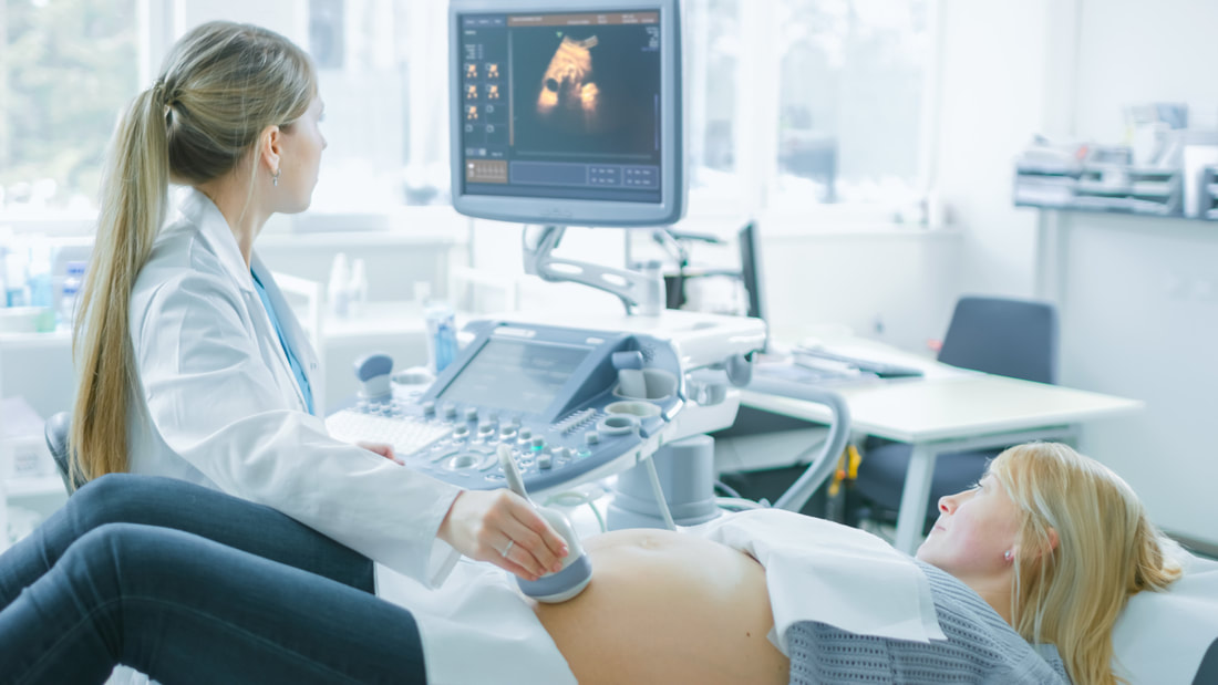
 A baby ultrasound, also called a sonogram or fetal ultrasound, is an imaging technique that uses sound waves to see your unborn child inside your womb. It is performed by a healthcare provider or medical technologist and can be used to detect and diagnose certain health problems and developmental abnormalities. It is a painless procedure and typically takes 30 minutes or less to complete. During your pregnancy ultrasound 3d procedure, the technician applies a gel to your abdomen (tummy) and moves a probe over the area. The sound waves reflect off the fetus and back to the transducer, which sends signals to a monitor that creates black-and-white images. You may be asked to lie on one side during the scan or to turn on both sides if images are needed from different positions. The basic anatomy scan - This type of ultrasound is done at about 12 weeks of pregnancy and uses sound waves to examine the baby's head, body and bones. It can help your doctor determine how far along you are and estimate your due date. It can also help identify any issues, such as multiple births or a placenta that is not growing normally. You will be asked to lay on your side and a thin gel will be applied to the surface of your tummy. This is to allow better contact between the scanner and your skin. Then the ultrasound probe is moved over your tummy and pictures are taken, which are sent instantly to a computer screen. The images are very clear and show a detailed view of your baby's body. At six weeks, an embryonic heart can be seen on the ultrasound screen. The technician can also view the fetus' brain and its internal organs, as well as the mother's uterus and the placenta. In some cases, a gynecologist can assess your baby's sex at this ultrasound by looking at the position of their genitals. However, this is not always possible. If you don't want to know, let the gynecologist know beforehand. If you are having a transvaginal ultrasound, the ultrasound hamilton practitioner will insert a small wand-like probe into your vaginal cavity. The wand transmits high-frequency sound waves that bounce off of the fetus and return to the device. The images are then converted to black-and-white pictures that the doctor can view on a screen. Your baby's size - This ultrasound is often done at the beginning of your third trimester to check that your baby is still on track for growth. The scan can also measure your baby's limb and finger lengths. If your baby is too short, this could indicate that they are not getting enough nourishment during the first few months of their life. During the ultrasound, your doctor can measure the width and length of your baby's rib cage and the bi-parietal diameter of the bones in your pelvic region. This information can be useful in predicting your baby's future weight and height. You can also choose to have a nuchal translucency scan, which is usually done at around 12 weeks of pregnancy. It can be combined with a blood test to check for chromosomal abnormalities. It can also help your doctor determine how likely it is that your baby will have a chromosomal disorder, such as Down syndrome. Here is more information on this topic: https://www.huffpost.com/archive/ca/entry/baby-ultrasound-this-photo-is-freaking-out-the-internet_n_8916674.
0 Comments
Leave a Reply. |
